Welcome to the ultimate guide to the wonders of ultrasound in the field of medicine. In this comprehensive article, we will take you on a journey through the fascinating world of this non-invasive and invaluable medical imaging tool. Whether you are a healthcare professional or simply curious about the advancements in medical technology, this guide aims to enlighten and educate.
Ultrasound has revolutionized the way medical practitioners diagnose and monitor various conditions, from pregnancy to cardiovascular diseases. With its ability to create real-time images of internal structures of the body, ultrasound plays a crucial role in guiding medical interventions and facilitating accurate diagnoses.
Throughout this guide, we will delve into the mechanics of ultrasound, explore its numerous applications across different medical specialties, and highlight its benefits and limitations. From obstetrics to orthopedics, ultrasound has become an indispensable tool in the healthcare industry, aiding in improved patient care and outcomes.
Join us as we unlock the marvels of ultrasound, providing you with a foundational understanding of this technology and its extensive contributions to the world of medicine. Get ready to be astounded by the power of sound waves and their endless possibilities in healthcare.
History of ultrasound in medicine
The journey of ultrasound in medicine began in the early 20th century, with the first significant developments occurring during World War II. Initially, ultrasound technology was primarily used for submarine detection and navigation. However, as scientists began to explore its potential in medical applications, the first clinical uses of ultrasound emerged in the late 1940s and early 1950s. Dr. Karl Dussik, an Austrian neurologist, is often credited with the first use of ultrasound in medicine when he utilized the technology to detect brain tumors using a technique called “ultrasonic transmission.” This pioneering work laid the groundwork for further advancements in the field.
As the technology evolved, ultrasound transitioned from research applications to clinical practice. The introduction of the A-mode (Amplitude mode) ultrasound in the 1950s allowed for the measurement of distances in the body, primarily for detecting abnormalities in organs. By the 1960s, B-mode (Brightness mode) ultrasound was developed, which enabled the visualization of tissues in a two-dimensional format. This advancement revolutionized diagnostic imaging, providing clinicians with a more detailed view of internal structures and leading to a broader range of applications in various medical fields.
Throughout the 1970s and 1980s, the development of real-time ultrasound imaging further transformed the landscape of medical diagnostics. The advent of Doppler ultrasound in the late 20th century provided a way to assess blood flow and cardiac function, enhancing the ability to diagnose cardiovascular conditions. With each technological advancement, ultrasound gained prominence as a safe, non-invasive, and effective imaging modality, becoming an essential tool in modern medicine.
How ultrasound works
Ultrasound operates on the principle of sound wave propagation. The process begins when a transducer, a specialized device that emits sound waves, is placed on the patient’s skin after applying a gel that enhances sound transmission. The transducer generates high-frequency sound waves, typically ranging from 2 to 18 megahertz, which travel through the body and reflect off internal structures such as organs, tissues, and fluids. These reflected sound waves are then captured by the transducer, which converts them into electrical signals.
The captured signals are processed by a computer to create real-time images displayed on a monitor. The varying densities and compositions of the tissues in the body result in different levels of echo, which determine the brightness and contrast of the images produced. For example, fluid-filled structures like the bladder appear dark on the ultrasound image, while solid tissues reflect more sound waves and appear brighter. This contrast allows healthcare providers to distinguish between healthy and abnormal tissues effectively.
One unique feature of ultrasound technology is its ability to provide dynamic imaging, allowing practitioners to visualize movement and function in real-time. This capability is particularly beneficial in assessing the heart’s function, observing blood flow, or guiding interventions in procedures such as biopsies. The non-invasive nature of ultrasound, combined with its ability to provide immediate results, makes it a valuable tool in a wide range of clinical settings.
Applications of ultrasound in medicine
Ultrasound is a versatile imaging modality with applications spanning numerous medical specialties. In obstetrics and gynecology, ultrasound is most commonly associated with monitoring pregnancy, allowing providers to visualize the developing fetus, assess gestational age, and identify potential complications. Routine ultrasounds help ensure the health and well-being of both the mother and the unborn child, making it an indispensable tool in prenatal care.
In the field of cardiology, echocardiography utilizes ultrasound to evaluate heart function and structure. This non-invasive technique enables cardiologists to assess conditions such as heart valve disorders, congenital heart defects, and cardiomyopathy. By visualizing the heart’s chambers and blood flow, echocardiography provides critical information that guides treatment decisions and management plans for patients with cardiovascular diseases.
Beyond obstetrics and cardiology, ultrasound plays a significant role in other specialties, including gastroenterology, urology, and orthopedics. In gastroenterology, ultrasound is used to evaluate abdominal organs, such as the liver and gallbladder, while in urology, it assists in assessing kidney stones and bladder conditions. In orthopedics, ultrasound aids in diagnosing musculoskeletal injuries and guiding injections or aspirating fluids from joints. The broad applicability of ultrasound highlights its importance in modern medical practice.
Benefits and limitations of ultrasound
The benefits of ultrasound in medicine are numerous, making it a favored imaging modality among healthcare providers. One of the most significant advantages is its non-invasive nature, allowing for the visualization of internal structures without the need for incisions or radiation exposure. This safety profile is particularly beneficial in vulnerable populations, such as pregnant women and children, where minimizing risk is paramount. Additionally, ultrasound is relatively cost-effective compared to other imaging modalities, such as MRI and CT scans, making it accessible for a wide range of patients.
Ultrasound also offers real-time imaging capabilities, which is crucial in various clinical situations. For instance, during a procedure, such as a needle aspiration or injection, ultrasound can guide the physician in real-time, ensuring accurate placement and reducing the risk of complications. Furthermore, the portability of ultrasound machines allows for bedside imaging, enhancing patient care in critical settings and emergency departments.
However, ultrasound does have its limitations. The quality of the images obtained can be affected by factors such as the patient’s body habitus, the presence of air or bone, and the operator’s experience. For example, in obese patients, the increased distance the sound waves must travel can diminish image quality. Additionally, ultrasound is not as effective in visualizing certain structures, such as the lungs or gastrointestinal tract, where gas can interfere with sound wave transmission. Despite these limitations, ongoing advancements in technology continue to enhance the capabilities of ultrasound, expanding its role in medical imaging.
Types of ultrasound imaging techniques
Ultrasound encompasses several imaging techniques, each tailored for specific clinical applications. The most common type is B-mode ultrasound, which provides two-dimensional images of internal structures. B-mode is widely used in obstetrics, cardiology, and abdominal imaging due to its ability to produce detailed images that help in diagnosing various conditions.
Doppler ultrasound is another critical technique that assesses blood flow and movement within the body. By measuring changes in frequency of the reflected sound waves, Doppler ultrasound can evaluate the velocity of blood flow in vessels and detect abnormalities such as blockages or valvular issues in the heart. This technique is invaluable in cardiology and vascular medicine, where understanding blood flow dynamics is crucial for diagnosis and treatment.
Additionally, there are specialized ultrasound techniques, such as 3D and 4D ultrasound, which provide three-dimensional images and real-time motion visualization, respectively. These advanced techniques are particularly beneficial in obstetrics, allowing for detailed assessments of fetal anatomy and well-being. Each type of ultrasound imaging technique plays a vital role in enhancing the diagnostic capabilities of healthcare providers, ensuring that patients receive accurate and timely evaluations.
Ultrasound in diagnostic medicine
In diagnostic medicine, ultrasound serves as a frontline imaging modality for various conditions and pathologies. One of its primary roles is in the evaluation of abdominal pain, where ultrasound can help identify issues such as gallstones, appendicitis, or liver disease. The ability to visualize solid organs and fluid collections in real-time allows clinicians to make informed decisions regarding further management or surgical intervention.
Moreover, ultrasound is essential in assessing the female reproductive system, providing valuable information in cases of pelvic pain, abnormal bleeding, or infertility. Transvaginal ultrasound offers enhanced visualization of the uterus and ovaries, allowing for the detection of conditions such as ovarian cysts, fibroids, or ectopic pregnancies. This targeted imaging approach is crucial for timely diagnosis and treatment planning.
In pediatrics, ultrasound is particularly beneficial due to its safety profile and non-invasive nature. Pediatricians use ultrasound to evaluate congenital anomalies, assess the status of the kidneys, and monitor the development of the hip joint in infants. The ability to perform these assessments without exposing children to ionizing radiation makes ultrasound an ideal choice in various diagnostic scenarios, ensuring the well-being of young patients.
Ultrasound in therapeutic medicine
Beyond diagnostics, ultrasound has therapeutic applications that contribute to patient care and treatment. One of the most common uses is in guided injections, where ultrasound technology helps physicians accurately place needles for corticosteroid injections in joints or soft tissues. This precision minimizes discomfort for the patient and maximizes the effectiveness of the treatment by ensuring that medication is delivered directly to the target site.
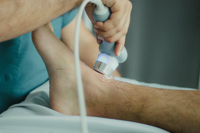
Additionally, ultrasound plays a role in physical therapy, where it is used for therapeutic ultrasound treatments. These treatments involve applying sound waves to tissues to promote healing, improve circulation, and reduce pain. The energy generated by ultrasound waves can enhance tissue repair processes, making it beneficial for conditions such as tendonitis, bursitis, and muscle strains.
Another innovative application of ultrasound in therapeutic medicine is in high-intensity focused ultrasound (HIFU), which is increasingly used in the treatment of certain tumors. HIFU utilizes focused ultrasound waves to generate heat, selectively destroying tumor cells while sparing surrounding healthy tissues. This non-invasive approach offers a promising alternative to traditional surgical methods, particularly for patients who are not candidates for surgery due to age or comorbidities.
Training and education in ultrasound
As ultrasound technology continues to advance, the demand for trained professionals skilled in ultrasound imaging is on the rise. Healthcare practitioners, including physicians, sonographers, and radiologists, require specialized training to effectively utilize ultrasound in clinical practice. Many medical schools and imaging programs now incorporate ultrasound education into their curricula, ensuring that future healthcare providers are well-versed in this essential imaging modality.
Certified ultrasound training programs provide hands-on experience and theoretical knowledge, covering topics such as anatomy, physiology, and image interpretation. Students learn to operate ultrasound machines, apply appropriate techniques, and understand the limitations and benefits of ultrasound imaging. Additionally, continuing education and workshops are crucial for professionals to stay updated with the latest advancements in ultrasound technology and best practices.
Professional organizations, such as the American Registry for Diagnostic Medical Sonography (ARDMS), offer certification exams for sonographers, ensuring that they meet established standards of competence. Certification not only enhances the credibility of professionals but also assures patients that they are receiving care from knowledgeable and skilled practitioners. As ultrasound becomes increasingly integral to patient care, ongoing training and education will remain essential for maintaining high standards in the field.
Future developments in ultrasound technology
The future of ultrasound technology is promising, with ongoing research and innovation poised to expand its applications and capabilities. One exciting development is the integration of artificial intelligence (AI) into ultrasound imaging. AI algorithms can assist in image analysis, enabling faster and more accurate interpretations of ultrasound results. By automating certain aspects of image evaluation, AI has the potential to reduce the workload on healthcare providers and improve diagnostic accuracy, leading to better patient outcomes.
Moreover, advancements in portable ultrasound devices are making this technology more accessible in various clinical settings. Handheld ultrasound machines are becoming increasingly sophisticated, allowing for bedside assessments in emergency situations or remote areas with limited access to traditional imaging facilities. This portability can significantly enhance patient care, providing rapid evaluations and facilitating timely interventions.
Additionally, researchers are exploring new ultrasound techniques, such as elastography, which assesses tissue stiffness and has implications for diagnosing liver fibrosis and tumors. The development of contrast-enhanced ultrasound is also gaining traction, allowing for improved visualization of blood flow and vascular structures. As technology continues to evolve, the potential for ultrasound to transform medical practice and enhance patient care remains vast and exciting.
