Are you ready to uncover the hidden secrets of X-ray technology? Look no further because this ultimate guide will take you on a captivating journey into the inner mysteries of this groundbreaking medical invention. X-ray technology has revolutionized the field of healthcare, allowing doctors to peer into the human body and diagnose injuries and illnesses with incredible precision.
In this comprehensive guide, we will delve into the history of X-rays, exploring how this incredible discovery transformed the way we understand and treat medical conditions. From Wilhelm Conrad Roentgen’s accidental discovery to the development of portable X-ray devices, we will explore the remarkable advancements that have taken place in this field.
But it doesn’t stop there. We will also unravel the science behind X-ray technology, explaining how it works and why it is such a valuable diagnostic tool. You will gain insight into the various applications of X-rays, ranging from dental examinations to detecting fractures and tumors.
Whether you’re a healthcare professional, a patient, or simply curious about the wonders of medical technology, this ultimate guide will satisfy your thirst for knowledge. Get ready for a captivating exploration of X-ray technology’s inner mysteries.
History of X-Ray Technology
The journey of X-ray technology began in the late 19th century, marked by a serendipitous discovery that would change the landscape of medicine forever. In 1895, German physicist Wilhelm Conrad Roentgen was conducting experiments with cathode rays when he stumbled upon a new form of radiation that could penetrate various materials. While testing his apparatus, he noticed that a nearby screen coated with barium platinocyanide began to glow, despite being shielded from the cathode rays. This unexpected phenomenon led Roentgen to realize he had discovered something extraordinary: a new type of ray that could produce images of the internal structures of objects, including the human body. He named this newfound radiation “X-rays,” with “X” symbolizing the unknown.
Roentgen’s groundbreaking work was published in a paper titled “On a New Kind of Rays,” which garnered immediate attention and acclaim. His first X-ray image, taken of his wife’s hand, showcased her wedding ring, captivating the world with the possibilities of this innovative technology. Within a year, X-ray machines began to emerge in medical practices across Europe and the United States. The ability to see inside the human body without invasive procedures transformed diagnostic medicine, setting the stage for a new era in healthcare.
As the 20th century progressed, the use of X-ray technology expanded rapidly. The development of more sophisticated machines and techniques, such as fluoroscopy, allowed for real-time imaging, further enhancing diagnostic capabilities. The application of X-rays during World War I for detecting injuries and foreign objects in soldiers underscored their importance in medical practice. By the mid-20th century, the introduction of the first portable X-ray machines revolutionized accessibility, enabling healthcare professionals to perform diagnostics in various settings, including field hospitals and remote areas.
How X-Rays Work
Understanding how X-rays work requires delving into the fundamental principles of physics. X-rays are a form of electromagnetic radiation, similar to visible light but with much shorter wavelengths and higher energy. When X-rays are produced, they emanate from a tube where electrons are accelerated and collide with a metal target, usually tungsten. This collision generates X-ray photons, which can penetrate through soft tissues but are absorbed by denser materials, such as bones. This differential absorption is key to producing the images that X-ray technology is known for.
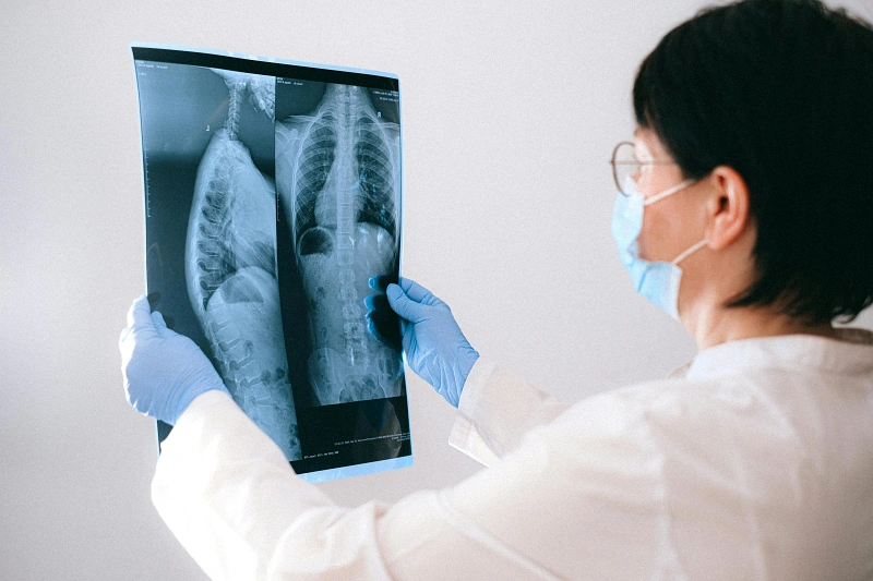
When an X-ray beam is directed at a patient, it passes through the body and strikes a detector or film on the opposite side. The varying degrees of absorption create a shadow-like effect, where denser structures such as bones appear white on the film, while softer tissues—like muscles and organs—appear in shades of gray. This contrast allows radiologists to interpret the images and identify any abnormalities, fractures, or diseases present. The process is quick, often taking only a few minutes, and is generally painless, making it a preferred diagnostic tool in many medical scenarios.
Moreover, advances in digital technology have revolutionized the way X-ray images are captured and interpreted. Digital X-ray systems utilize electronic sensors instead of traditional film, enabling immediate image acquisition and processing. This not only enhances image quality but also reduces the radiation exposure to patients. Furthermore, digital images can be easily stored, shared, and analyzed using sophisticated software, facilitating more accurate diagnoses and improved patient care.
Types of X-Ray Imaging
X-ray technology encompasses various imaging modalities, each designed to serve specific diagnostic purposes. Traditional radiography is the most common form, providing standard images of bones and certain soft tissues. This technique is widely used in emergency departments for evaluating fractures, joint dislocations, and other orthopedic issues. The straightforward nature of conventional X-ray imaging makes it an invaluable tool for initial assessments and routine examinations in clinical practice.
Fluoroscopy is another vital type of X-ray imaging that allows real-time visualization of internal structures and functions. By continuously emitting X-rays, fluoroscopy enables healthcare professionals to observe dynamic processes, such as the movement of the gastrointestinal tract during a swallow study or the flow of blood through vessels. This real-time imaging is instrumental in guiding various procedures, such as catheter placements and orthopedic surgeries, enhancing the precision and efficacy of interventions.
Computed Tomography (CT) scanning represents a more advanced X-ray modality that combines multiple X-ray images taken from different angles to create cross-sectional views of the body. CT scans provide detailed and intricate images, allowing for the assessment of complex anatomical structures and the detection of diseases at earlier stages. While CT imaging exposes patients to higher levels of radiation compared to traditional X-rays, its diagnostic power has made it an essential tool in oncology, trauma, and many other medical fields.
Applications of X-Ray Technology in Medicine
X-ray technology has a wide array of applications in the medical field, significantly enhancing diagnostic capabilities across various specialties. One of the most common uses is in orthopedics, where X-rays are utilized to evaluate fractures, joint conditions, and bone diseases. The clear images produced by X-rays allow physicians to assess the extent of injuries, plan treatment strategies, and monitor healing progress over time. This is crucial in ensuring optimal recovery for patients with musculoskeletal injuries.
In dentistry, X-ray imaging plays an essential role in diagnosis and treatment planning. Dental X-rays, such as bitewings and panoramic radiographs, provide vital information about tooth structure, root health, and the presence of cavities or infections. Dentists rely on these images to detect issues that may not be visible during a clinical examination, allowing for timely interventions and improved oral health outcomes. The use of digital dental X-rays has further streamlined the process, enabling faster image acquisition and reduced radiation exposure.
X-ray technology is also integral to oncology, where it aids in the detection and monitoring of tumors. Radiologists utilize X-ray imaging, including mammography for breast cancer screening and chest X-rays for lung cancer evaluation, to identify suspicious masses and assess their progression over time. Additionally, X-ray guidance is employed during certain therapeutic procedures, such as radiation therapy, ensuring that cancerous tissues are targeted accurately while minimizing damage to surrounding healthy tissues.
Advancements in X-Ray Technology
The evolution of X-ray technology has been marked by significant advancements that have improved its effectiveness and safety. One of the most notable innovations has been the development of digital X-ray systems, which have largely replaced traditional film-based imaging. Digital systems offer numerous advantages, including immediate image availability, enhanced image quality, and the ability to manipulate images for better visualization. This shift not only streamlines workflows in medical facilities but also reduces the environmental impact associated with film processing.
Another major advancement has been the introduction of advanced imaging techniques, such as dual-energy X-ray absorptiometry (DEXA). This technique is primarily used for assessing bone density, aiding in the diagnosis of osteoporosis and other bone-related conditions. DEXA scans provide precise measurements of bone mineral density, allowing for early detection and intervention in patients at risk for fractures. The accuracy and non-invasive nature of DEXA have made it a standard procedure in many healthcare settings.
Artificial intelligence (AI) is poised to transform the landscape of X-ray technology further. AI algorithms can analyze vast amounts of imaging data, identifying patterns and anomalies that may escape the human eye. This capability enhances diagnostic accuracy and efficiency, reducing the burden on radiologists and expediting patient care. Additionally, AI-driven tools can assist in predicting patient outcomes and guiding treatment decisions, making X-ray technology not only a diagnostic tool but also a pivotal player in personalized medicine.
Safety Considerations in X-Ray Imaging
While X-ray technology is an invaluable diagnostic tool, it is essential to consider the associated safety implications, particularly regarding radiation exposure. X-rays involve the use of ionizing radiation, which, if not properly managed, can pose potential health risks. To mitigate these risks, healthcare providers adhere to the principle of ALARA (As Low As Reasonably Achievable), which emphasizes minimizing radiation exposure while still obtaining necessary diagnostic information.
Patients are often advised to inform their healthcare providers of any previous X-ray examinations, especially if they have undergone multiple scans within a short period. This information helps radiologists make informed decisions about the necessity of additional imaging and explore alternative diagnostic methods when appropriate. In some cases, advanced imaging techniques, such as MRI or ultrasound, may provide sufficient information without the use of ionizing radiation.
Protective measures are also implemented during X-ray procedures to enhance patient safety. Lead aprons and shields are commonly used to protect sensitive organs and tissues from unnecessary exposure. Additionally, advancements in technology, such as digital imaging and automated dose control systems, have contributed to reducing radiation doses while maintaining image quality. Ongoing research into safer imaging techniques and protocols continues to play a vital role in ensuring patient safety in the realm of X-ray technology.
Future of X-Ray Technology
The future of X-ray technology is poised for exciting developments that promise to enhance diagnostic capabilities and improve patient outcomes. One area of ongoing research is the enhancement of image quality through the use of novel materials and detection systems. Innovations in detector technology, such as photon-counting detectors, are expected to provide higher resolution images with lower radiation doses, making X-ray examinations safer and more effective.
Moreover, the integration of machine learning and AI into radiology is anticipated to revolutionize image interpretation. AI algorithms are being trained to recognize specific patterns associated with various medical conditions, enabling faster and more accurate diagnoses. This technology has the potential to assist radiologists in prioritizing cases based on urgency, optimizing workflow, and ultimately improving patient care. As AI continues to evolve, its role in X-ray imaging will likely expand, leading to more personalized and efficient healthcare solutions.
Telemedicine and remote consultations are also expected to impact the future of X-ray technology. As healthcare increasingly embraces digital platforms, the ability to share X-ray images electronically allows for remote interpretation by specialists, regardless of geographic location. This advancement is particularly beneficial in rural or underserved areas where access to expert radiologists may be limited. By facilitating timely consultations and second opinions, telemedicine will enhance patient care, making X-ray technology an even more powerful tool in modern healthcare.
X-Ray Technology in Non-Medical Fields
Beyond its prominent role in medicine, X-ray technology has found applications in various non-medical fields, showcasing its versatility and effectiveness. One notable application is in the field of security, where X-ray machines are commonly used for scanning luggage and cargo at airports and border crossings. These machines allow security personnel to inspect the contents of bags without opening them, identifying potential threats such as weapons or explosives. The ability to see through materials has made X-ray technology a cornerstone in ensuring public safety.
X-ray imaging is also utilized in industrial applications, particularly in non-destructive testing (NDT). This technique enables engineers and quality control inspectors to examine the integrity of materials and components without causing damage. Industries such as aerospace, automotive, and construction rely on X-ray technology to detect structural flaws, corrosion, and other defects in critical components. By ensuring the reliability and safety of materials, X-ray technology plays a vital role in maintaining quality standards across various industries.
Additionally, X-ray technology has applications in art and archaeology. Conservators and researchers use X-ray imaging to analyze artworks and historical artifacts, gaining insights into their construction, materials, and condition. For example, X-ray fluorescence (XRF) can identify the elemental composition of pigments, revealing information about an artist’s techniques and materials. In archaeology, X-rays can help uncover hidden layers and structures within artifacts, providing valuable context for historical studies. These applications highlight the broader significance of X-ray technology beyond the medical realm, demonstrating its impact on various fields.
Conclusion: The Impact of X-Ray Technology on Society
The advent of X-ray technology has had a profound impact on society, revolutionizing the way we diagnose and treat medical conditions. From its humble beginnings with Wilhelm Conrad Roentgen’s accidental discovery to its current status as an essential tool in modern medicine, X-rays have transformed healthcare practices and improved patient outcomes. The ability to visualize the internal structures of the human body has not only enhanced diagnostic accuracy but has also facilitated the development of innovative treatment approaches.
Moreover, the advancements in X-ray technology continue to shape the future of healthcare, with ongoing research and development focused on improving safety, efficiency, and accuracy. As we embrace digital transformation, the integration of AI and telemedicine is set to further enhance the capabilities of X-ray imaging, ensuring that patients receive timely and effective care. The cross-disciplinary applications of X-ray technology in security, industry, and the arts underscore its versatility and importance in addressing various societal needs.
In summary, X-ray technology represents a remarkable achievement in scientific progress, with far-reaching implications for medicine and beyond. As we continue to unveil the inner mysteries of X-ray technology, its potential to enhance our understanding of the human body, improve diagnostic practices, and safeguard society remains limitless. The legacy of X-rays will undoubtedly continue to evolve, enriching the fields of healthcare, industry, and research for generations to come.


