Ultrasound examinations are commonly used in medical practice to provide a clear, real-time view inside the body. Many patients encounter this test during routine check-ups, pregnancy, or when investigating specific symptoms. While ultrasound is a valuable tool, it’s important to understand both its strengths and its limitations. This article will guide you through how ultrasound works, its most frequent uses, what it cannot detect, and when your healthcare provider may recommend additional tests.
How ultrasound works
Ultrasound, sometimes called sonography, is a medical imaging technique that uses high-frequency sound waves to create images of the inside of the body. Unlike X-rays or CT scans, it does not involve radiation, making it a safe option for a wide range of patients, including pregnant women and children.
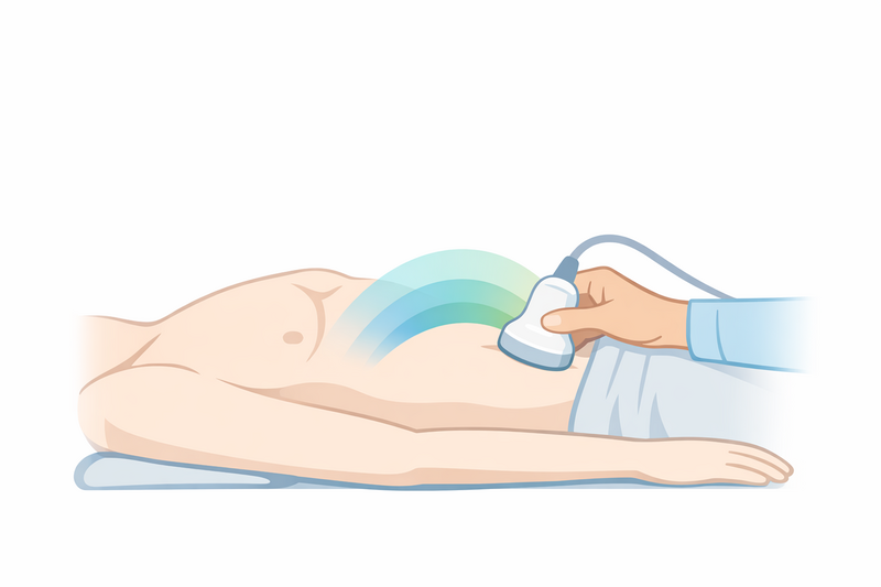
The examination is usually performed by a radiologist or a specially trained technician called a sonographer. During the procedure, a small handheld device called a transducer is placed on the skin, usually with a layer of gel to improve contact. The transducer sends sound waves into the body, which bounce off tissues and organs. These echoes are picked up by the transducer and translated into images on a monitor.
- Non-invasive: No needles or incisions are required.
- Painless: Most patients feel only mild pressure from the transducer.
- Real-time images: Results are available immediately during the scan.
Ultrasound can be performed on many parts of the body. The quality of the images depends on the area being examined, the skill of the operator, and the patient’s individual characteristics.
Common uses
Ultrasound is a versatile imaging method, and its applications cover many areas of medicine. Some of the most common uses include:
- Pregnancy: Monitoring fetal development, checking the baby’s position, and assessing the placenta or amniotic fluid.
- Abdominal organs: Examining the liver, gallbladder, spleen, pancreas, and kidneys for abnormalities such as cysts, stones, or inflammation.
- Pelvic organs: Assessing the uterus, ovaries, bladder, and prostate for structural changes or growths.
- Blood vessels: Doppler ultrasound can measure blood flow and detect blockages or narrowing in arteries and veins.
- Thyroid and neck: Identifying nodules, enlargement, or other structural changes in the thyroid gland or surrounding tissues.
- Musculoskeletal system: Visualising muscles, tendons, and joints to detect tears, fluid collections, or inflammation.
Doctors may also use ultrasound to guide certain medical procedures, such as biopsies or fluid drainage, because the real-time images help improve accuracy and safety.
Advantages in specific patient groups
Because ultrasound does not use radiation, it is especially suitable for:
- Pregnant women, for monitoring the health of both mother and baby
- Children, who may be more sensitive to radiation exposure
- Patients who require frequent imaging, such as those with chronic conditions
Ultrasound is often the first imaging test ordered because it is quick, safe, and widely available.
What ultrasound cannot detect
Although ultrasound is a powerful diagnostic tool, it does have important limitations. Understanding what ultrasound cannot show is essential for setting realistic expectations and for recognising when additional tests may be needed.
- Air-filled structures: Ultrasound waves do not pass well through air. This means that organs filled with air, such as the lungs or intestines, are not well visualised.
- Bone detail: While ultrasound can show the surface of bones, it cannot penetrate them. It is not suitable for detailed imaging of the brain (in adults) or for evaluating bone marrow or fractures deep inside the bone.
- Small or deep-lying abnormalities: Very small tumors, especially those located deep within the body or behind bone or air-filled organs, may be missed.
- Obesity or excess tissue: In patients with a high body mass index (BMI), the quality of ultrasound images can be reduced, making it harder to see certain organs or detect abnormalities.
- Microscopic changes: Ultrasound shows structures, not cells. It cannot detect microscopic changes, such as early cancer development or minor tissue abnormalities.
In addition, certain conditions can be difficult to distinguish on ultrasound. For example, some benign and malignant growths may appear similar, which is why further tests are sometimes necessary to confirm a diagnosis.
Examples of limitations
- Ultrasound is not reliable for detecting early-stage cancers in most organs, as small tumors may not show up clearly.
- It cannot accurately assess the inside of the intestines or lungs due to the presence of gas or air.
- It is not a substitute for mammography in breast cancer screening, although it may be used as an additional test.
Your doctor will take these limitations into account when recommending an ultrasound or interpreting the results.
When further tests are needed
After an ultrasound, your doctor may sometimes recommend additional tests. This can happen for several reasons, such as:
- The ultrasound results are unclear or inconclusive.
- The images show a possible abnormality that needs to be investigated further.
- Your symptoms do not match the ultrasound findings.
Additional tests may include:
- CT scans: Use X-rays to produce detailed cross-sectional images, especially useful for bones, lungs, and complex organs.
- MRI scans: Use magnetic fields to generate detailed images of soft tissues, nerves, and organs deep inside the body.
- Mammography: Specialised X-rays for breast tissue, often used alongside ultrasound for breast concerns.
- Biopsy: Taking a small sample of tissue for laboratory analysis, usually guided by imaging methods.
- Endoscopy: Direct visual inspection of internal organs using a flexible tube with a camera.
Your doctor will select the most appropriate follow-up tests based on your individual situation, medical history, and the findings on the ultrasound. It’s important to remember that needing further tests does not always mean something serious is wrong; often, it is simply a precaution to ensure accurate diagnosis and the best possible care.
If you are scheduled for further imaging, such as an MRI scan, you may find it helpful to read more about what to expect during an MRI and what doctors look for in these scans to better prepare yourself for the process.
Working with your healthcare team
If you have questions about your ultrasound results or why further tests are recommended, do not hesitate to ask your doctor. Open communication can help you understand the reasons behind each step in your care and reduce unnecessary worry.
In summary, ultrasound is a safe, non-invasive, and widely used imaging technique that plays a key role in modern medicine. While it can reveal a great deal about the body’s internal structures, it does have limitations. Your healthcare provider may use ultrasound as part of a broader diagnostic approach, combining it with other tests when necessary to provide you with the most accurate and complete picture of your health.