What Is a PET Scan?
A Positron Emission Tomography (PET) scan is a specialized imaging technique used in medicine to visualize metabolic processes in the body. Unlike traditional imaging methods like X-rays or CT scans, which show structural details, PET scans provide insights into the functioning of tissues and organs. This makes it a valuable tool for diagnosing and managing conditions such as cancer, heart disease, and neurological disorders.
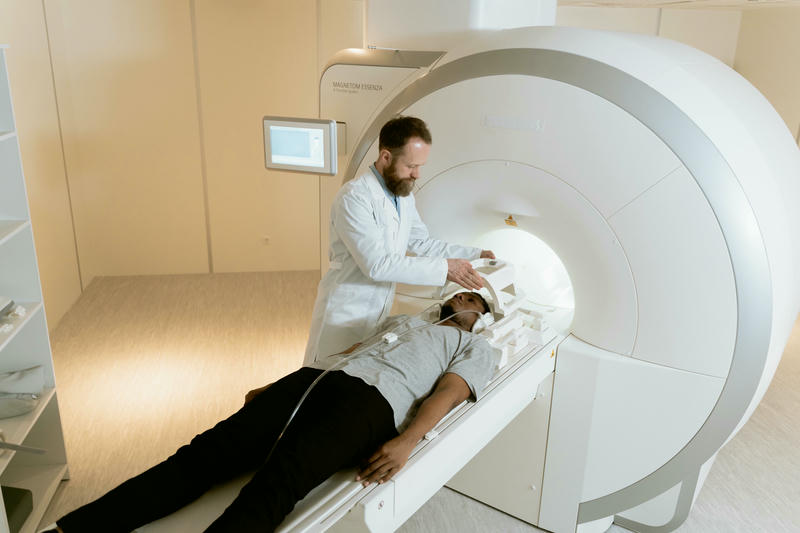
During a PET scan, a small amount of a radioactive substance, called a tracer, is introduced into the body. The tracer accumulates in areas with high metabolic activity, and the PET scanner detects this activity to produce detailed images. This process enables doctors to detect abnormalities that might not be visible through other imaging modalities.
The Technology Behind PET Scans
The technology behind a PET scan involves the interplay of physics, chemistry, and advanced computing. The key component is the tracer, which is a molecule labeled with a radioactive isotope. A commonly used tracer is fluorodeoxyglucose (FDG), which mimics glucose, the body’s primary energy source.
After the tracer is injected into the bloodstream, it travels to tissues where it undergoes decay, emitting positrons (positively charged particles). When these positrons encounter electrons, they annihilate each other, producing gamma rays. The PET scanner detects these gamma rays and uses algorithms to reconstruct a 3D image of the body, highlighting areas of high metabolic activity.
PET scanners are often combined with CT or MRI machines to create hybrid imaging systems, such as PET/CT or PET/MRI, which provide both metabolic and structural information. This combination enhances diagnostic accuracy and allows doctors to pinpoint abnormalities with greater precision.
How PET Scan Results Are Evaluated
The results of a PET scan are interpreted by a radiologist or nuclear medicine specialist. After the scan, the specialist examines the images to identify areas of abnormal tracer uptake. These areas are typically color-coded, with brighter colors indicating higher levels of metabolic activity.
For instance:
- In cancer diagnosis, increased uptake of the tracer may indicate tumor growth.
- In heart disease, reduced uptake in certain regions of the heart can suggest poor blood flow or damage.
- In neurological disorders, abnormal patterns of tracer distribution may help identify conditions like Alzheimer’s disease or epilepsy.
The findings from a PET scan are often compared with results from other imaging tests or clinical evaluations to provide a comprehensive diagnosis. The detailed report is shared with the patient’s physician, who explains the results and outlines potential next steps.
Potential Risks of PET Scans
PET scans are generally safe, but like any medical procedure, they come with some risks. The radioactive tracer used in the scan involves exposure to a low level of radiation. For most patients, this exposure is minimal and within safe limits. However, it may not be suitable for pregnant women or individuals with certain medical conditions.
Common side effects may include:
- A mild allergic reaction to the tracer.
- Temporary discomfort at the injection site.
- Rarely, feelings of dizziness or nausea.
Before the scan, patients are asked about their medical history, allergies, and current medications to minimize risks. It is important to follow all instructions provided by the healthcare team, such as fasting before the scan, to ensure accurate results and reduce potential side effects.
How Long Have PET Scans Been Used in Medical Practice?
PET technology has been in use since the 1970s, but its widespread adoption in clinical practice began in the 1990s. Over the decades, advancements in tracer development, scanner technology, and imaging software have significantly improved its capabilities and accessibility.
Today, PET scans are a standard diagnostic tool in many medical fields, including oncology, cardiology, and neurology. They have revolutionized the way diseases are diagnosed and treated, allowing for earlier detection and more personalized treatment plans.
The Future of PET Scans
The future of PET technology is promising, with ongoing research focused on expanding its applications and improving its precision. Key developments include:
- New tracers designed to target specific diseases, enabling more precise diagnosis and monitoring.
- Integration with artificial intelligence (AI) to enhance image analysis and reduce interpretation time.
- Improved scanner designs that offer higher resolution and faster imaging, reducing scan times and patient discomfort.
Additionally, PET imaging is being explored for use in emerging fields like immunotherapy and personalized medicine. Researchers are investigating how PET can guide treatments by visualizing the effectiveness of therapies in real time.
As technology advances, PET scans are likely to become even more integral to medical care, offering patients and doctors powerful tools for understanding and managing complex health conditions.
Summary
Positron Emission Tomography (PET) scans are a cutting-edge imaging technology that helps doctors visualize metabolic activity in the body. By using a radioactive tracer and advanced scanners, PET provides critical insights into diseases like cancer, heart conditions, and neurological disorders. While the procedure involves minimal risks, its benefits in early diagnosis and treatment planning are immense. With decades of proven use in medical practice and ongoing advancements in technology, PET scans continue to play a vital role in modern healthcare, with even greater potential for the future.
