Welcome to the definitive guide to understanding Computed Tomography (CT) scan. In this article, we will unravel the mysteries of this powerful medical imaging technique and explain how it plays a vital role in diagnosing and treating various conditions.
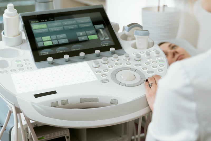
CT scan, also known as a CAT scan, uses a series of X-ray images taken from different angles to create cross-sectional images of the body. It provides detailed insight into the internal structures, helping healthcare professionals detect and evaluate a wide range of medical conditions, from injuries to cancers.
Whether you’ve recently been prescribed a CT scan or are simply curious about how it works, this guide will walk you through the entire process, covering everything from the basics of CT scanning to its potential risks and benefits.
By the end of this article, you’ll have a clear understanding of what to expect, how to prepare, and why CT scanning is such a valuable tool in modern medicine.
So, let’s get started and crack the code of Computed Tomography!
How does a CT scan work?
Computed Tomography (CT) scans operate by employing a sophisticated combination of X-ray technology and computer processing to create detailed images of the body’s internal structures. During a CT scan, a patient lies on a movable table that slides into a large, circular machine called a gantry. The gantry houses an X-ray tube and a series of detectors that rotate around the patient. As the X-ray tube emits a narrow beam of radiation, the detectors capture the X-rays that pass through the body, recording varying levels of absorption based on the density of the tissues encountered.
The data collected during the scan is then transmitted to a computer, which processes the information to generate cross-sectional images, or slices, of the body. These slices can be viewed in multiple planes, giving healthcare providers a comprehensive view of the organs and tissues. The ability to visualize structures in various orientations allows for better assessment and diagnosis of potential health issues. CT scans can produce images in 3D format, adding another layer of detail that can aid in surgical planning and other medical interventions.
CT technology has advanced over the years, leading to improvements in image quality and reduced scan times. Modern CT scanners can perform high-resolution scans with less radiation exposure compared to older models. Innovations such as multi-slice CT, which captures multiple slices simultaneously, enhance the speed and efficiency of the imaging process. This advancement not only improves patient comfort but also enables quicker diagnoses, allowing for timely medical decisions and interventions.
Types of CT scans
There are several types of CT scans, each designed for specific diagnostic purposes. One of the most common is the standard CT scan, which provides detailed images of various body parts, including the head, chest, abdomen, and pelvis. This type of scan is particularly useful for detecting tumors, injuries, and internal bleeding. Additionally, specialized CT scans, such as a CT angiography, focus on blood vessels and can help diagnose conditions like aneurysms or blockages.
Another variation is the high-resolution CT scan, typically used for assessing lung diseases. This type of scan captures more detailed images of the lung structures, aiding in the diagnosis of conditions such as interstitial lung disease or emphysema. Furthermore, CT colonography, also known as virtual colonoscopy, is a minimally invasive procedure used to screen for colorectal cancer. It utilizes CT technology to create images of the colon and rectum, allowing for the detection of polyps or other abnormalities.
Some CT machines are equipped for specific examinations, such as CT-guided biopsies, where imaging helps guide a needle to obtain tissue samples from specific locations within the body. This precision increases the accuracy of diagnoses while minimizing discomfort for the patient. Overall, the diversity of CT scan types allows healthcare providers to tailor imaging techniques to the individual needs of patients, enhancing diagnostic capabilities across various medical fields.
Advantages and limitations of CT scans
CT scans offer numerous advantages, making them a vital tool in modern diagnostics. One of the primary benefits is their speed; a CT scan can be completed in a matter of minutes, providing rapid results that are critical in emergency situations. This quick turnaround is especially crucial for trauma cases where immediate imaging can determine life-saving interventions. Additionally, CT scans are highly detailed, producing clear images that help in the accurate assessment of complex structures, allowing for better diagnosis and treatment planning.
Another significant advantage of CT scans is their ability to visualize not just bones, but also soft tissues, blood vessels, and organs, all in one imaging session. This comprehensive imaging capability is beneficial for a variety of medical conditions, including cancer, vascular diseases, and internal injuries. Moreover, advancements in technology have led to reduced radiation exposure, making modern CT scans safer than those from earlier years. Techniques such as iterative reconstruction algorithms further enhance image quality while minimizing dose, allowing for safer imaging practices.
However, despite their advantages, CT scans also come with limitations. One primary concern is the exposure to ionizing radiation, which, while reduced in modern scans, can still accumulate over time for patients undergoing multiple scans. This raises questions about the long-term risks, especially in younger patients or those requiring repeated imaging. Additionally, CT scans may not always provide sufficient information for certain conditions, necessitating further imaging studies or alternative diagnostic methods, such as MRI or ultrasound.
Common uses of CT scans in medical diagnosis
CT scans are employed in a wide array of medical diagnoses, making them indispensable in contemporary healthcare. One of the most common applications is in oncology, where CT imaging helps identify tumors, determine their size, and assess whether cancer has spread to surrounding tissues or lymph nodes. This information is critical for staging cancer and formulating effective treatment plans. Furthermore, CT scans are often used to monitor the effectiveness of treatment over time, allowing for adjustments based on the tumor’s response.
Another significant use of CT scans is in the evaluation of traumatic injuries. In cases of accidents or falls, CT scans can quickly reveal internal bleeding, fractures, or organ damage. This rapid assessment enables healthcare providers to make swift decisions regarding surgical interventions or other necessary treatments. In emergency departments, CT scans play a pivotal role in triaging patients with head injuries, abdominal pain, or chest pain, ensuring timely and appropriate care.
Additionally, CT scans are valuable in diagnosing various medical conditions beyond cancer and trauma. For instance, they are often used to investigate diseases of the lungs, such as pneumonia, pulmonary embolism, or chronic obstructive pulmonary disease (COPD). They can also aid in diagnosing gastrointestinal issues, such as appendicitis, diverticulitis, and bowel obstructions. The versatility of CT imaging allows it to serve as a cornerstone in the diagnostic process across multiple specialties, including cardiology, neurology, and orthopedics.
Preparing for a CT scan
Preparation for a CT scan can vary depending on the type of scan being performed and the area of the body being examined. Typically, patients will receive specific instructions from their healthcare provider in advance of the procedure. One common requirement is fasting for a certain period before the scan, especially for abdominal or pelvic CT scans. This helps ensure that the images are as clear as possible by reducing the presence of food or fluid in the digestive tract that could obscure the view.
Patients may also need to inform their healthcare provider of any medications they are taking or any allergies they may have, particularly to contrast materials used during the scan. A contrast agent, often administered intravenuously, enhances the visibility of certain structures in the images. While most people tolerate contrast materials well, some individuals may experience allergic reactions. In such cases, alternative imaging options may be considered.
In addition, it is important for patients to wear comfortable clothing and avoid any metal accessories, such as jewelry or belts, which can interfere with the imaging process. In certain instances, patients may be asked to change into a hospital gown. Understanding the preparation requirements can help alleviate anxiety and ensure a smooth scanning process, leading to more accurate diagnostic results.
What to expect during a CT scan procedure
During a CT scan procedure, patients can expect a relatively straightforward and non-invasive experience. After checking in at the imaging department, a technologist will guide the patient through the process, providing instructions and answering any questions. Once in the scanning room, the patient will be positioned on a padded table, which will slide into the CT machine. It is important to remain as still as possible during the scan to avoid blurring the images.
The duration of a CT scan can vary based on the complexity of the exam, but most scans take only a few minutes. Patients may hear a series of clicking or whirring sounds as the machine rotates and captures images. Depending on the specific scan, the technologist may ask the patient to hold their breath for a few seconds during certain parts of the imaging to enhance the quality of the results.
If a contrast agent is used, it may be administered through an IV line, and patients might experience a warm sensation or a metallic taste in their mouth as the contrast material flows through their body. These sensations are typically temporary and resolve quickly. After the scan is complete, patients can usually resume their normal activities immediately, although some may be advised to drink extra fluids if contrast was used to help eliminate it from their system.
Interpreting CT scan results
Once the CT scan is completed, the images are sent to a radiologist, a medical doctor specialized in interpreting medical images. The radiologist carefully examines the scans for any signs of abnormalities, such as tumors, fractures, or signs of inflammation. They assess the images from various angles and may compare them to previous scans if available. The detailed analysis allows for a comprehensive evaluation of the area of concern.
After interpreting the results, the radiologist compiles a report summarizing their findings and provides recommendations or insights based on the images. This report is then sent to the referring healthcare provider, who will discuss the results with the patient. In some cases, the provider may recommend further imaging studies, additional tests, or specific treatments based on the findings of the CT scan.
It is important for patients to understand that while CT scans provide valuable information, they are just one component of the diagnostic process. The results must be interpreted in conjunction with a patient’s medical history, symptoms, and other diagnostic tests. Open communication between the patient and healthcare provider is essential to ensure that the findings are understood and that appropriate steps are taken for further evaluation or treatment.
Risks and safety considerations of CT scans
While CT scans are invaluable diagnostic tools, they also carry certain risks and safety considerations that patients should be aware of. One of the primary concerns is the exposure to ionizing radiation. Although modern CT technology has significantly reduced radiation doses, the cumulative effects of multiple scans can still pose a potential risk, particularly for vulnerable populations such as children or individuals requiring frequent imaging. Healthcare providers strive to follow the principle of “as low as reasonably achievable” (ALARA) when it comes to radiation exposure.
In addition to radiation concerns, patients may experience allergic reactions to contrast materials used during some CT scans. Although severe allergic reactions are rare, mild reactions such as itching or rash may occur. Patients with a history of allergies or kidney problems should inform their healthcare provider prior to the procedure to ensure appropriate precautions are taken. Pre-medication with antihistamines or corticosteroids may be necessary for those with known allergies to contrast agents.
Furthermore, it is essential for pregnant women to discuss the necessity of a CT scan with their healthcare provider. The potential risks to the developing fetus must be carefully weighed against the benefits of the imaging study. In many cases, alternative imaging modalities that do not involve radiation, such as ultrasound or MRI, may be considered to reduce potential risks. Overall, understanding these risks helps patients make informed decisions regarding their healthcare and the use of imaging technologies.
Conclusion: The importance of CT scans in modern healthcare
In conclusion, Computed Tomography (CT) scans play a critical role in modern healthcare, providing essential insights that aid in the diagnosis and treatment of various medical conditions. Their ability to generate detailed cross-sectional images allows healthcare professionals to visualize internal structures with remarkable clarity, facilitating accurate diagnoses and timely interventions. From oncology to trauma care, CT scans have revolutionized the way medical professionals assess and manage patient health.
As technology advances, the safety and effectiveness of CT scans continue to improve. The introduction of lower radiation doses, faster scan times, and enhanced image quality has made CT imaging more accessible and reliable. Despite potential risks, the benefits of CT scans often outweigh the downsides, particularly when they lead to early detection of life-threatening conditions or guide critical treatment decisions.
Ultimately, understanding CT scans empowers patients to engage in their healthcare actively. By knowing what to expect before, during, and after the procedure, individuals can approach their imaging studies with confidence and clarity. As a cornerstone of diagnostic medicine, CT scans will undoubtedly remain a vital tool in the ongoing quest for improved patient outcomes and enhanced healthcare quality.
