Key points
- CT is faster and is often preferred for bone, lungs, acute bleeding, and urgent internal problems.
- MRI gives more detailed views of soft tissues such as the brain, spinal cord, joints, ligaments, and pelvic organs.
- CT uses ionising radiation, while MRI does not, but MRI has important safety limits with some metal implants and devices.
- The choice depends on the body part, suspected condition, urgency, contrast needs, and patient safety factors.
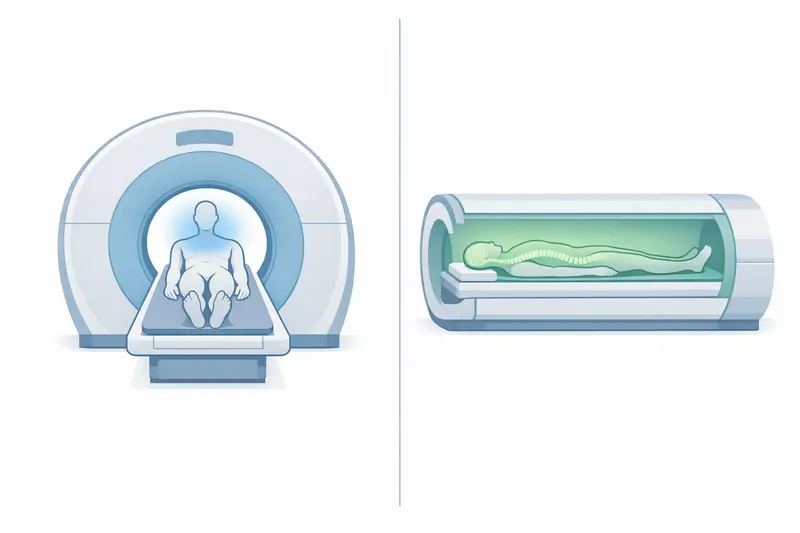
CT and MRI scans are both widely used imaging tests, but they do not do the same job. A CT scan uses X-rays to create cross-sectional images quickly, while an MRI uses a strong magnetic field and radio waves to show soft tissues in much greater detail. For a broader overview of medical imaging procedures and how they fit into diagnosis and treatment, see Examinations and Treatments Explained for Patients.
For patients, the difference matters because the best scan depends on the body part, the suspected problem, speed, safety factors, and what information the doctor needs. This guide explains what each scan shows, when one may be preferred over the other, and the main safety and practical differences.
What a CT scan shows compared to an MRI scan
A CT scan is often better for showing bone, lungs, acute bleeding, and many urgent internal problems. It produces detailed cross-sectional images very quickly, which is why it is commonly used in emergency settings. For example, CT may be used to assess suspected head injury, stroke bleeding, chest infection complications, kidney stones, bowel perforation, or fractures that are difficult to see clearly on a standard X-ray.
An MRI scan is usually better for showing soft tissues. This includes the brain, spinal cord, nerves, muscles, ligaments, cartilage, pelvic organs, and many internal organs when fine tissue detail is needed. MRI is often used to evaluate conditions such as a torn knee ligament, a slipped disc, multiple sclerosis, liver lesions, prostate abnormalities, or suspected problems affecting the brain and spinal cord.
How the images differ
CT images are very good at showing differences in density, especially between air, bone, fluid, and fresh blood. MRI images are more sensitive to differences within soft tissue, which can make inflammation, oedema, tumours, ligament injuries, and changes in the brain or spinal cord easier to detect.
Contrast agents may be used in either test to improve visibility. In CT, iodine-based contrast is commonly used to show blood vessels, inflammation, tumours, or organ injury. In MRI, gadolinium-based contrast may help highlight abnormal tissue activity, blood supply, or breakdown of normal tissue barriers, such as in some brain conditions.
- CT often shows best: fractures, lung disease, acute internal bleeding, kidney stones, trauma-related injuries
- MRI often shows best: ligaments, cartilage, spinal discs, brain tissue, nerves, bone marrow, pelvic soft tissues
- Both can assess: tumours, organ disease, infection, inflammation, and blood vessels, but one may be more informative depending on the clinical question
When doctors choose a CT scan instead of an MRI
A doctor may choose CT when speed is important. CT scans are usually much faster than MRI and are often available in emergency departments around the clock. In acute situations, a scan that can be completed within minutes may be more useful than a slower test, even if MRI may later provide more detailed soft-tissue information.
CT is commonly preferred in situations such as:
- major trauma after a road accident or fall
- suspected bleeding in the brain
- assessment of the chest in severe breathing symptoms or suspected pulmonary embolism
- suspected kidney stones
- evaluation of complex fractures
- urgent abdominal pain, especially when bowel obstruction, perforation, appendicitis complications, or organ injury are concerns
Practical reasons CT may be chosen
CT is also easier for some patients to tolerate. The scan is quick, the machine is more open than most MRI scanners, and body movement is less likely to spoil the images. This can matter for patients with severe pain, agitation, confusion, or breathing difficulty.
Another reason is access to equipment or medical devices. Some implants, monitoring equipment, or emergency support devices may limit the use of MRI. CT is often more practical when intensive monitoring is needed or when metallic implants have not yet been fully checked for MRI safety.
In cancer care, CT is frequently used to assess the chest, abdomen, and pelvis because it is fast, widely available, and useful for mapping disease spread in organs such as the lungs, liver, or bowel. It may also be used to guide biopsies or drain collections.
When doctors prefer an MRI over a CT scan
MRI is often preferred when the main question involves soft-tissue detail rather than speed. If the doctor needs to look closely at the brain, spinal cord, joints, ligaments, pelvic organs, or bone marrow, MRI may provide much more useful information than CT.
Common examples include:
- persistent back pain with suspected disc prolapse or nerve compression
- knee, shoulder, or ankle injuries involving ligaments, tendons, or cartilage
- suspected brain tumours, multiple sclerosis, or subtle stroke changes
- spinal cord compression
- liver, uterus, ovaries, or prostate assessment when more tissue characterisation is needed
- investigation of bone marrow disorders or hidden fractures not clearly seen on X-ray or CT
When avoiding radiation matters
MRI may also be preferred when avoiding ionising radiation is important, especially in younger patients or when repeated imaging is likely. For example, a patient with a chronic neurological condition, inflammatory bowel disease, or repeated joint problems may need several scans over time. In these situations, avoiding repeated CT radiation can be an important consideration. For those preparing for an MRI, it’s helpful to know what to expect and how to prepare for the scan.
Why MRI can answer different questions
MRI does not simply produce a sharper version of a CT image. It provides different types of tissue information. Different MRI sequences can highlight fluid, inflammation, fat, blood products, nerve pathways, or tissue perfusion. This is why MRI may detect abnormalities that are subtle or invisible on CT, particularly in the brain, spinal cord, and joints.
Risks and safety differences between CT and MRI
The most important safety difference is that CT uses ionising radiation, while MRI does not. A single CT scan is often medically justified when the information is important, but radiation exposure is still considered carefully, especially in children, younger adults, and patients who may need repeated scans.
MRI avoids radiation, but it has its own safety concerns because of the strong magnetic field. Metal implants, fragments, or devices may pose a risk or affect image quality. Some pacemakers are MRI-compatible, but this must be checked in advance. A patient should always inform staff about implants, surgical clips, cochlear implants, metal injuries to the eye, or implanted pumps and stimulators.
Contrast-related risks
Both scans may involve contrast, and each has different considerations.
- CT contrast: usually iodine-based; may rarely cause allergic reactions and may require extra caution in patients with significantly reduced kidney function
- MRI contrast: usually gadolinium-based; is often well tolerated, but may also require caution in severe kidney disease
If contrast is planned, staff may ask about previous contrast reactions, asthma, kidney disease, diabetes medication, and recent blood test results. Not every CT or MRI scan requires contrast.
Claustrophobia and comfort
MRI is more likely than CT to cause discomfort related to noise, time, and the enclosed scanner tunnel. The patient usually needs to lie still for longer, sometimes 20 to 45 minutes or more depending on the body area and number of sequences. CT is usually much shorter, often completed within a few minutes.
For patients with claustrophobia, severe pain, or difficulty lying flat, CT may be easier to complete. Some MRI departments can offer coping strategies, headphones, communication systems, or in some cases mild sedation, depending on local practice and the clinical situation.
Limitations of CT scans and MRI scans
Neither scan is “better” in every situation. Each has limitations, and the most useful test depends on the clinical question.
Limitations of CT
- uses radiation
- shows soft tissues less clearly than MRI in many parts of the body
- may miss subtle brain, spinal cord, ligament, cartilage, or marrow abnormalities
- iodine contrast may not be suitable for every patient
For example, CT can identify a major stroke bleed quickly, but it may be less sensitive than MRI for some early ischaemic strokes or small lesions in the posterior brain. CT can also show a joint well in terms of bone structure, but it may not define tendon or ligament injury as well as MRI.
Limitations of MRI
- takes longer and is less practical in some emergencies
- is more sensitive to patient movement
- may not be possible with some implants or metal fragments
- can be difficult for patients with claustrophobia or severe pain
- is often less useful than CT for lung detail, calcifications, and some acute fractures
MRI is excellent for many soft-tissue problems, but it is not always the first choice for acute trauma, suspected bowel perforation, or rapid assessment of unstable patients. In these situations, CT may offer the right balance of speed and diagnostic value.
In practice, doctors choose between CT and MRI based on the suspected diagnosis, the body area involved, how urgently the result is needed, whether contrast is necessary, the patient’s medical history, and whether radiation or MRI safety issues are relevant. Sometimes both scans are used at different stages because they provide complementary information.