Are you looking for a comprehensive guide on mammography for early detection? Look no further. In this article, we will provide you with everything you need to know about this essential screening tool. Whether you are a woman approaching the recommended age for your first mammogram or someone who wants to learn more about the procedure, we’ve got you covered.

Mammograms play a vital role in detecting breast cancer in its early stages, leading to higher survival rates and a better chance of successful treatment. But what exactly is a mammogram? How often should you get one? Are there any risks involved? We’ll answer these questions and more, equipping you with the knowledge to make informed decisions about your healthcare.
Our goal is to empower you with the information you need to understand the importance of mammography and its role in early detection. By the end of this article, you’ll be well-informed and prepared for your next mammogram. So let’s dive in and learn everything you need to know about mammography for early detection.
What is mammography?
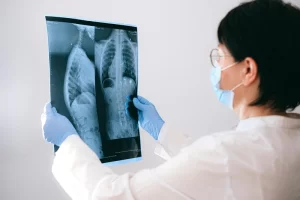
Mammography is a specialized medical imaging technique that uses low-energy X-rays to examine the human breast. The primary purpose of mammography is to detect early signs of breast cancer, long before physical symptoms develop. This screening method is crucial for women, especially those over the age of 40, as it provides a non-invasive way to identify abnormalities in breast tissue. The images produced by mammography, known as mammograms, allow radiologists to identify any suspicious areas that may warrant further investigation.
There are two main types of mammography: screening and diagnostic. Screening mammograms are performed on women who do not exhibit any symptoms of breast cancer. These are typically conducted annually or biennially, depending on a woman’s age and risk factors. In contrast, diagnostic mammograms are performed when a woman presents symptoms such as a lump or unusual breast changes. These examinations provide a more detailed view of the breast tissue, helping healthcare providers assess any areas of concern more accurately.
The technology behind mammography has evolved significantly over the years, transitioning from traditional film-based systems to digital mammography. Digital mammograms offer improved image quality and allow for enhanced manipulation of images, making it easier for radiologists to analyze breast tissue. Additionally, breast tomosynthesis, or 3D mammography, is becoming increasingly popular as it provides a three-dimensional view of the breast, reducing the chance of false positives and improving the detection of small tumors.
Importance of early detection in breast cancer
Early detection of breast cancer is vital as it significantly increases the chances of successful treatment and survival. When breast cancer is caught in its early stages, it is often localized and has not yet spread to nearby lymph nodes or other parts of the body. This localized stage is far more manageable and typically requires less aggressive treatment options, such as lumpectomy or radiation therapy, rather than more extensive procedures like mastectomy or chemotherapy.
The statistics speak for themselves: according to the American Cancer Society, women diagnosed with localized breast cancer have a five-year survival rate of nearly 99%. This statistic highlights the importance of routine screenings and raises awareness about the need for women to prioritize their breast health. By participating in regular mammography screenings, women can take proactive steps toward detecting any potential issues early on, ultimately saving lives.
Moreover, early detection not only impacts survival rates but also affects the quality of life for those diagnosed with breast cancer. Women who undergo treatment for early-stage breast cancer often experience fewer side effects and a quicker recovery compared to those diagnosed at more advanced stages. This means that not only are their chances of survival higher, but they can also return to their normal lives sooner, with less physical and emotional disruption.
Benefits of mammography screening
Mammography screening provides numerous benefits, making it an essential tool in the fight against breast cancer. One of the primary advantages is the potential for early detection, which we have already discussed. By identifying breast cancer at its earliest stage, women can receive prompt treatment, improving their prognosis and reducing the overall burden of the disease.
Another significant benefit of mammography is its ability to detect cancers that may not be palpable during a physical examination. Many tumors can be too small to feel, and mammograms can reveal these tiny abnormalities long before they become noticeable. This early detection increases the likelihood of catching cancer when it is most treatable, ultimately leading to better outcomes for patients.
Additionally, mammography has been shown to reduce breast cancer mortality rates. Studies have demonstrated that regular screening mammograms can lower the risk of dying from breast cancer by approximately 15-30%, depending on various factors such as age and population demographics. This statistic underscores the critical role of mammography in promoting public health and encouraging women to take charge of their breast health through regular screenings.
Mammography guidelines and recommendations
Mammography guidelines vary depending on the organization, but several key recommendations are widely accepted. The American College of Radiology (ACR) and the Radiological Society of North America (RSNA) recommend that women begin annual screening mammograms at age 40. However, some organizations, like the U.S . Preventive Services Task Force (USPSTF), suggest that women aged 50 to 74 undergo screening every two years, while those under 50 should consult with their healthcare provider to make an informed decision based on their individual risk factors.
Women with a family history of breast cancer or other risk factors may need to start screening earlier. For those with a significantly increased risk, the guidelines recommend discussing personalized screening plans with a healthcare provider. This may include starting mammograms before the age of 40 or incorporating additional imaging modalities, such as MRI, into their screening routine.
It is essential for women to stay informed about any updates to screening guidelines and recommendations. As research continues to evolve, healthcare organizations may adjust their recommendations to improve early detection and patient outcomes. Regular consultations with healthcare providers can help women understand their specific risks and develop a tailored screening plan that meets their individual needs.
Types of mammography exams
There are primarily two types of mammography exams: screening mammograms and diagnostic mammograms. Screening mammograms are the standard procedure recommended for women who do not have any symptoms of breast cancer. These exams are typically conducted annually or biennially and involve taking two X-ray images of each breast from different angles. The primary goal of a screening mammogram is to identify any early signs of breast cancer or abnormalities that may require further investigation.
Diagnostic mammograms, on the other hand, are utilized when a woman presents symptoms or when a screening mammogram indicates a potential concern. This type of mammogram provides a more detailed examination of specific areas of the breast. The imaging process is similar to a screening mammogram, but the radiologist may take additional images to focus on any areas of interest. Diagnostic mammograms are often accompanied by a clinical examination and may be recommended after a physical exam reveals a lump or other concerning changes in breast tissue.
In recent years, 3D mammography, or breast tomosynthesis, has gained popularity as an advanced imaging technique. This method creates a three-dimensional picture of the breast, allowing radiologists to examine the tissue layer by layer. The benefits of 3D mammography include improved detection rates of small tumors and a reduction in false-positive results. Women may be able to choose between traditional 2D mammography and 3D mammography, depending on their individual preferences and the availability of technology at their screening facility.
How to prepare for a mammogram
Preparing for a mammogram involves several simple steps to ensure the process goes smoothly and produces the most accurate results. Firstly, it is advisable to schedule your mammogram for a time when your breasts are least likely to be tender. For many women, this means avoiding the week before their menstrual period, as hormonal changes can lead to increased sensitivity. Choosing the right timing can make the experience more comfortable.
On the day of the mammogram, it is essential to wear a two-piece outfit. This allows for easy removal of the top clothing without needing to disrobe completely. Additionally, women should refrain from using any deodorant, lotion, or perfume on the day of the exam, as these products can interfere with the imaging process. Some deodorants contain metallic particles that can show up on the mammogram, leading to potential confusion during interpretation.
Finally, it is vital to inform the radiologic technologist about any previous breast surgeries, biopsies, or history of breast cancer. Bringing previous mammograms, if available, can help the radiologist compare current images to past results. Making sure to communicate any concerns or symptoms you may have, such as lumps or changes in breast appearance, will ensure the technologist is aware and can make the necessary adjustments during the exam.
What to expect during a mammogram
When you arrive for your mammogram appointment, you will be greeted by a radiologic technologist who will guide you through the process. After completing any necessary paperwork, you will be taken to a private changing area where you will be asked to change into a gown. It is normal to feel a bit anxious, but understanding the procedure can help alleviate some of that anxiety.
During the mammogram, you will stand in front of the mammography machine. The technologist will position your breast on a flat surface and will gently compress it with a clear plastic paddle. This compression is crucial for obtaining clear images as it spreads out the breast tissue and reduces the amount of radiation needed. While the compression may feel uncomfortable, it typically lasts only a few seconds. You may be asked to hold your breath briefly while the images are taken, which helps eliminate motion blur.
The entire process usually takes about 15 to 30 minutes, including the time spent changing and positioning. After the mammogram is complete, you can resume your normal activities. The images will be reviewed by a radiologist, who will analyze the results for any abnormalities. Depending on the findings, you may receive your results within a few days or weeks, and your healthcare provider will discuss the next steps if further evaluation is necessary.
Understanding mammogram results
Interpreting mammogram results is an essential aspect of the screening process. After your mammogram, the images will be analyzed by a radiologist who will look for any signs of abnormalities, such as masses, calcifications, or changes in breast tissue density. The results are typically categorized using the Breast Imaging Reporting and Data System (BI-RADS), which assigns a score from 0 to 6 based on the findings.
A BI-RADS score of 0 indicates that additional imaging is needed, while scores from 1 to 3 suggest that the results are normal or benign. A score of 4 or 5 indicates that there is a suspicious finding that may require further investigation, such as a biopsy. A score of 6 means that the patient has a known biopsy-proven malignancy. Understanding these categories can help women grasp the significance of their results and the potential next steps.
It is essential to remember that a mammogram is not a definitive test for breast cancer. Abnormal findings may not always indicate cancer, and many women with suspicious results do not have breast cancer. In some cases, additional imaging, such as ultrasound or MRI, may be recommended to gain a clearer understanding of the findings. If a biopsy is necessary, it will provide definitive answers regarding the presence or absence of cancer.
Common concerns and misconceptions about mammography
Many women have concerns and misconceptions regarding mammography, which can deter them from scheduling regular screenings. One common fear is that mammograms are painful. While some discomfort may occur during the compression of the breast, it is typically brief and manageable. Understanding that this compression is necessary for obtaining clear images can help alleviate anxiety surrounding the procedure.
Another misconception is that mammograms expose women to high levels of radiation. In reality, the amount of radiation used in a mammogram is minimal and considered safe. The benefits of early detection far outweigh the risks associated with exposure to radiation, especially when considering the potential consequences of undetected breast cancer. Healthcare providers emphasize the importance of routine screenings as a proactive measure for breast health.
Lastly, some women may believe that mammograms are not necessary if they have no family history of breast cancer. However, breast cancer can occur in women with no familial links. In fact, most women diagnosed with breast cancer do not have a family history of the disease. Regular mammography screenings are essential for all women, regardless of their family history, as they provide the best chance for early detection and successful treatment.
Conclusion: Taking control of your breast health through mammography
Taking control of your breast health through mammography is an empowering step that every woman should consider. Understanding the importance of early detection, the benefits of regular screenings, and the procedures involved can help dispel fears and misconceptions surrounding mammography. By prioritizing routine mammograms, women can significantly improve their chances of detecting breast cancer in its earliest stages, ultimately leading to better treatment outcomes and survival rates.
As you navigate your healthcare journey, remember to consult with your healthcare provider about your individual risk factors and discuss the appropriate timing for your mammograms. Staying informed about the latest guidelines and recommendations will help you make educated decisions about your breast health.
In summary, mammography is a vital tool in the fight against breast cancer. By embracing regular screenings, women can take proactive measures to safeguard their health and well-being. So, schedule your next mammogram, encourage your friends and family to do the same, and take an active role in protecting your breast health for years to come.

