Welcome to the comprehensive guide on Magnetic Resonance Imaging (MRI), where we unlock the secrets behind this groundbreaking technology. In this article, we will delve into the intricate workings of MRI, providing you with a clear understanding of how it revolutionizes medical diagnostics.

With its ability to capture detailed images of the body’s internal structures, MRI has become an invaluable tool in modern medicine. By utilizing a powerful magnetic field and radio waves, MRI creates highly detailed cross-sectional images that aid in the diagnosis and monitoring of various health conditions.
Throughout this guide, we will explore the fundamentals of MRI, including its history, principles, and the different types of MRI scanners available today. You will discover how MRI is used to detect and diagnose a wide range of medical issues, from detecting tumors and evaluating joint injuries to diagnosing neurological disorders and exploring the complexities of the brain.
Whether you are a healthcare professional or simply curious about this remarkable technology, this guide will provide you with a comprehensive overview of MRI and its essential role in modern healthcare. So, let’s embark on this fascinating journey of unlocking the secrets of Magnetic Resonance Imaging.
How does MRI work?
Magnetic Resonance Imaging (MRI) operates on the principles of nuclear magnetic resonance, a phenomenon that was first discovered in the 1940s. At its core, MRI utilizes powerful magnets and radio waves to generate detailed images of the body’s internal structures. When a patient enters the MRI scanner, the strong magnetic field aligns the hydrogen atoms present in the body’s tissues. Hydrogen is abundant in the human body, mainly due to water content, making it an ideal target for imaging.
Once the hydrogen atoms are aligned, the MRI machine emits radiofrequency pulses that excite these atoms, causing them to emit signals as they return to their original alignment. These signals are detected by the MRI machine and are processed by a computer to produce high-resolution images. The resulting images can be viewed in various planes, giving healthcare professionals a comprehensive look at the anatomy and any abnormalities present. This detailed imaging capability is what makes MRI such a powerful diagnostic tool in modern medicine.
MRI technology has evolved significantly since its inception. Early machines produced images that were less detailed and took considerably longer to generate. Advances in technology, such as the development of stronger magnets and more sophisticated computer algorithms, have resulted in faster scan times and improved image quality. Additionally, functional MRI (fMRI) has emerged, allowing for the observation of brain activity by measuring changes in blood flow, further expanding the applications of this remarkable technology.
In summary, the working of MRI hinges on the interaction between magnetic fields, radio waves, and hydrogen atoms within the body. This unique combination allows for the creation of detailed images that are invaluable for diagnosing a wide array of medical conditions. As we delve deeper into the advantages and limitations of MRI, we will better understand its role in modern healthcare and how it compares to other imaging modalities.
Advantages and limitations of MRI
MRI boasts numerous advantages that make it a preferred imaging technique in various clinical scenarios. One of the primary benefits is its ability to produce high-resolution images with exceptional contrast, particularly of soft tissues. This characteristic allows for the accurate visualization of organs, muscles, and other structures that may be difficult to assess with other imaging modalities like X-rays or CT scans. Furthermore, MRI does not use ionizing radiation, making it a safer option for patients, especially for those requiring multiple imaging studies over time.
Another significant advantage of MRI is its versatility. It can be used to examine many body parts, including the brain, spine, joints, and abdomen. Additionally, advanced MRI techniques, such as diffusion-weighted imaging and spectroscopy, provide insights into specific conditions like tumors, strokes, and neurodegenerative diseases. The ability to capture images in multiple planes and create 3D reconstructions further enhances its diagnostic capabilities.
However, MRI is not without its limitations. One of the most notable drawbacks is the cost associated with the procedure. MRI machines are expensive to purchase and maintain, which can lead to higher costs for patients and healthcare providers. Additionally, MRI scans can take longer than other imaging tests, often requiring patients to remain still for extended periods, which may be challenging for some individuals.
Moreover, certain patients may not be suitable candidates for an MRI scan. Those with implanted medical devices, such as pacemakers or cochlear implants, may face risks due to the strong magnetic fields used in MRI. Additionally, individuals who experience claustrophobia may find it uncomfortable to undergo an MRI, as the machines can be enclosed and noisy. Therefore, while MRI offers many advantages, it is essential to weigh these against its limitations in specific clinical contexts.
Types of MRI scans
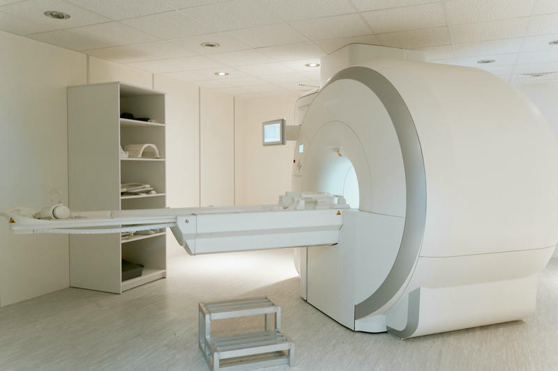
MRI technology has diversified, giving rise to several types of scans tailored for specific diagnostic needs. One of the most common types is the standard MRI scan, which provides detailed images of various anatomical structures. This scan is typically performed with the patient lying on a table that slides into the MRI machine. Depending on the region being examined, specific sequences may be employed to optimize the visibility of different tissues.
Another important type is functional MRI (fMRI), which assesses brain activity by measuring blood flow changes. This technique is particularly useful in neurological research and clinical settings, as it helps identify areas of the brain responsible for specific functions. fMRI is often employed pre-surgically to map critical brain regions in patients undergoing neurosurgery, ensuring that vital areas are preserved during procedures.
In addition, there are specialized MRI techniques, such as contrast-enhanced MRI and cardiac MRI. Contrast-enhanced MRI involves the administration of a contrast agent, typically gadolinium, to enhance the visibility of blood vessels and certain tissues, making it easier to identify abnormalities such as tumors or inflammation. Cardiac MRI, on the other hand, focuses on the heart’s structure and function, providing valuable insights into conditions like heart disease, cardiomyopathy, and congenital heart defects. These various types of MRI scans highlight the technology’s adaptability and its critical role in diagnosing a wide range of medical conditions.
Preparing for an MRI scan
Preparation for an MRI scan is essential to ensure the procedure’s success and the patient’s safety. Prior to the appointment, patients are typically asked to provide a detailed medical history, including any existing medical conditions, allergies, and medications they are taking. It is crucial for the healthcare provider to know about any implanted devices, such as pacemakers, metal plates, or other foreign objects, as these may pose risks during the scan.
Patients may also be instructed to remove any metal objects, including jewelry, watches, and hairpins, before entering the MRI room. This precaution is necessary because metal can interfere with the magnetic field and affect image quality. Additionally, some MRI scans may require patients to fast for a specific period before the procedure, particularly if a contrast agent will be used. Following the healthcare provider’s instructions carefully can help minimize the chances of complications and ensure optimal imaging results.
For individuals who experience anxiety or claustrophobia, discussing these concerns with the healthcare team ahead of time is crucial. Sedatives may be offered to help relax anxious patients, and alternative imaging methods, such as open MRI machines, may be considered for those who cannot tolerate traditional closed MRI scanners. By adequately preparing for the MRI scan, patients can alleviate any apprehension and contribute to a smoother experience during the procedure.
What to expect during an MRI scan
When patients arrive for an MRI scan, they will be greeted by a healthcare professional who will explain the procedure and answer any questions. The patient will be asked to change into a gown and will need to remove any remaining metal objects. Once ready, the patient will lie on a padded table that slides into the MRI machine. Depending on the type of scan, the patient may be positioned in various ways to ensure the area of interest is adequately imaged.
During the scan, it is essential for patients to remain still, as movement can blur the images. The MRI machine will emit loud noises, such as banging or thumping sounds, which are normal and result from the rapid switching of magnets. Patients are often provided with earplugs or headphones to help reduce the noise level. While the scan can take anywhere from 15 minutes to over an hour, depending on the complexity of the imaging being performed, the healthcare team will keep patients informed throughout the process.
In some cases, a contrast agent may be administered through an intravenous (IV) line to enhance the images further. The injection is typically quick and may cause a brief sensation of warmth. After the scan is complete, patients can resume their normal activities, as there are usually no restrictions following an MRI. The images will be reviewed by a radiologist, who will provide a report to the referring physician for further evaluation and discussion with the patient.
Common uses of MRI in medical diagnosis
MRI technology is widely used in various medical fields due to its ability to provide detailed images of soft tissues, organs, and structures within the body. One of the most common applications is in the diagnosis of neurological conditions. MRI is the gold standard for detecting brain tumors, multiple sclerosis, strokes, and other abnormalities in the central nervous system. By providing high-resolution images of the brain and spinal cord, MRI helps neurologists formulate accurate diagnoses and treatment plans.
In orthopedics, MRI plays a critical role in evaluating joint injuries, such as tears in ligaments and cartilage. It is particularly valuable for diagnosing conditions like anterior cruciate ligament (ACL) tears in the knee and rotator cuff injuries in the shoulder. The detailed images produced by MRI allow for better assessment of the extent of the injury, guiding the choice of treatment, whether conservative management or surgical intervention.
Additionally, MRI is used in oncology to assess tumors in various body parts, including the breast, liver, and prostate. It helps determine the size, location, and extent of cancerous growths, which is crucial for staging and planning treatment. Breast MRI, in particular, is often used as an adjunct to mammography for high-risk patients or to evaluate suspicious findings. The versatility of MRI makes it an indispensable tool in modern medical diagnostics across multiple specialties.
Risks and safety considerations of MRI
While MRI is generally considered a safe imaging technique, certain risks and safety considerations must be kept in mind. One primary concern is the presence of metallic implants or foreign objects within the body. Patients with devices such as pacemakers, certain types of stents, or metal fragments may be at risk during an MRI due to the strong magnetic field. It is essential for patients to disclose any such devices to their healthcare provider before the scan to determine if it is safe to proceed.
Another consideration is the use of contrast agents, which are typically used to enhance image quality. Although gadolinium-based contrast agents are generally safe, they can cause allergic reactions in some individuals. Additionally, patients with kidney dysfunction may be at risk for nephrogenic systemic fibrosis (NSF) after receiving gadolinium. Therefore, it is crucial for healthcare providers to assess kidney function prior to administering contrast agents.
Claustrophobia is another concern for some patients undergoing MRI scans, particularly in traditional closed MRI machines. For those who experience anxiety in confined spaces, discussing these feelings with the healthcare team is vital. Alternative options, such as open MRI machines or sedation, may be recommended to ensure a comfortable experience. By addressing these risks and considerations, healthcare providers can help ensure patient safety and comfort throughout the MRI process.
Future advancements in MRI technology
As technology continues to evolve, the field of MRI is poised for significant advancements that promise to enhance its capabilities and applications. One area of ongoing research is the development of higher-field-strength MRI systems, which can provide even greater image resolution and speed. These advanced systems have the potential to improve diagnostic accuracy and reduce scan times, making MRI more efficient for both patients and healthcare providers.
Another exciting development is the integration of artificial intelligence (AI) and machine learning into MRI interpretation. These technologies can assist radiologists in analyzing images more quickly and accurately by identifying patterns and anomalies that may be easily overlooked. AI algorithms can also streamline workflow processes, allowing for faster diagnosis and improved patient outcomes.
Additionally, there is ongoing research into new MRI techniques, such as hyperpolarized MRI, which enhances the signal from specific molecules, enabling the visualization of metabolic processes in real time. This emerging technology could revolutionize the diagnosis and monitoring of various conditions, including cancer and metabolic disorders. As MRI technology continues to advance, it holds the promise of providing even more comprehensive insights into human health and disease.
Conclusion
Magnetic Resonance Imaging (MRI) has undoubtedly transformed the landscape of medical diagnostics, offering unparalleled insights into the human body without the risks associated with ionizing radiation. From its intricate workings to its vast applications across various medical fields, MRI stands as a testament to the power of technology in advancing healthcare. Understanding the principles, benefits, limitations, and future directions of MRI is essential for both healthcare professionals and patients alike.
As we have explored in this guide, MRI serves a crucial role in diagnosing neurological disorders, musculoskeletal injuries, and various cancers, among other conditions. Its ability to provide detailed images of soft tissues makes it an invaluable tool for clinicians, enabling them to make informed decisions regarding patient care. However, it is equally important to recognize the risks associated with MRI, particularly concerning metal implants and the use of contrast agents, ensuring patient safety remains a priority.
Looking ahead, the future of MRI is bright, with ongoing advancements promising to enhance its capabilities and applications. As researchers continue to explore innovative techniques and integrate artificial intelligence into the imaging process, we can anticipate even more precise and efficient diagnostic tools. By unlocking the secrets of Magnetic Resonance Imaging, we pave the way for better healthcare outcomes and a deeper understanding of the complexities of the human body.


