Key points
- CT uses ionising radiation and carries a small long-term risk that matters more with repeated scans and in younger patients.
- MRI does not use radiation, but metal implants, electronic devices, and claustrophobia may affect safety or feasibility.
- Ultrasound is generally the safest common imaging test, though its main limitation is that it cannot answer every clinical question.
- Imaging is safest when the expected diagnostic benefit outweighs the risks and the test is matched to the clinical problem.
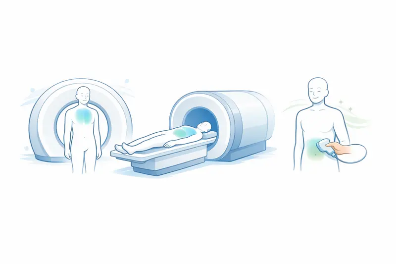
Most medical imaging tests are considered safe when used for the right reason and with the right precautions. The main differences lie in the type of energy they use: CT scans use ionising radiation, MRI uses strong magnets and radio waves, and ultrasound uses sound waves. For a broader look at how different examinations are chosen and what patients can expect, see Examinations and Treatments Explained for Patients.
For patients, the practical questions are usually straightforward: how much risk is involved, who may need extra care, and how doctors decide whether a scan is worth doing. This article explains the safety profile of common imaging tests, when the risks matter more, and how those risks are reduced in everyday medical practice.
Are CT scans safe and what are the radiation risks?
CT scans are widely used and can be very important in both urgent and non-urgent care. They produce detailed cross-sectional images and are often used to assess problems such as suspected stroke, major injury, appendicitis, kidney stones, lung disease, cancer staging, or internal bleeding.
The main safety concern with CT is exposure to ionising radiation. Unlike MRI and ultrasound, CT uses X-rays. Radiation from a single CT scan usually remains within a low to moderate medical range, but it is higher than with a standard plain X-ray.
What the radiation risk actually means
The concern is not that a CT scan causes immediate harm in most people. The issue is a small increase in lifetime cancer risk, especially if a person has repeated scans over time or is exposed at a younger age. This risk depends on several factors, including:
- the body part being scanned
- the scan settings and technique used
- age, with children and younger adults generally being more sensitive
- how many scans have already been done
For example, a CT scan of the head usually involves less radiation than a CT scan of the abdomen and pelvis. A patient with recurrent kidney stones or inflammatory bowel disease may accumulate more exposure over the years than someone having a single emergency scan after an accident.
When the benefit is greater than the risk
In many situations, the benefit of CT clearly outweighs the radiation risk. This is often the case when a quick and accurate answer is needed, such as:
- checking for bleeding after a head injury
- looking for a blood clot in the lungs
- assessing severe abdominal pain
- guiding cancer diagnosis or urgent treatment decisions
In these settings, missing a serious condition may be far more harmful than the radiation from the scan itself. For a detailed look at how CT and MRI differ in specific situations, you can read the key differences between CT and MRI scans.
Special points about contrast dye
Some CT scans also use contrast material, often iodine-based, to make blood vessels and organs easier to see. This does not create radiation risk, but it has its own safety considerations. A small number of patients may have an allergic-type reaction, and contrast may be more relevant in people with significant kidney impairment.
That is why staff may ask about previous contrast reactions, asthma, kidney disease, diabetes medication such as metformin, and recent blood test results.
Are MRI scans safe for all patients?
MRI does not use ionising radiation, so it does not carry the same radiation-related cancer concern as CT. It is often chosen to image the brain, spinal cord, joints, muscles, liver, pelvis, and some heart conditions, especially when detailed soft tissue imaging is needed.
However, MRI is not automatically safe for every patient in every circumstance because it uses a very strong magnetic field.
Who may need extra screening before MRI
The main safety issue is whether a person has metal or electronic devices in or on the body that could move, heat up, malfunction, or distort the images. Important examples include:
- certain pacemakers or implanted defibrillators
- some older aneurysm clips
- cochlear implants
- certain neurostimulators or infusion pumps
- metal fragments, especially in the eye
Many modern implants are MRI-conditional rather than completely prohibited, meaning MRI may still be possible under specific conditions such as particular scanner settings or body positions. This is why detailed safety screening is essential before the scan.
Common concerns during the scan
Even when MRI is medically safe, the experience can be difficult for some patients. The scanner is noisy, the space is enclosed, and the person often needs to lie still for 15 to 45 minutes or longer, depending on the examination. Claustrophobia, pain when lying flat, and an inability to stay still can sometimes prevent a successful scan.
In some cases, ear protection, extra communication, mild sedation, or an open MRI system may help. Young children and some adults with severe anxiety or movement disorders may need sedation or anaesthesia.
What about MRI contrast agents
Some MRI examinations use gadolinium-based contrast agents. These are generally well tolerated, but they are not risk-free. They may be used to assess inflammation, tumours, blood vessels, or active disease in tissues.
Extra caution is usually needed in people with severe kidney failure because of a rare but serious complication called nephrogenic systemic fibrosis. Allergic reactions can also occur, although they are less common than with some CT contrast agents.
Are ultrasound examinations considered safe?
Ultrasound is generally considered the safest of the common imaging methods because it uses sound waves rather than ionising radiation. It is widely used in pregnancy, abdominal imaging, pelvic examinations, vascular studies, and assessment of soft tissues such as the thyroid, testes, breast, or muscles.
For most patients, ultrasound has no known harmful effects when used appropriately by trained professionals for a clear medical reason.
Why ultrasound is often preferred
Ultrasound is often chosen first when doctors want an imaging test that is quick and avoids radiation. Examples include:
- checking for gallstones in a person with right upper abdominal pain
- examining pelvic organs in someone with abnormal bleeding
- looking for deep vein thrombosis in a swollen leg
- monitoring fetal growth and position during pregnancy
It can also be repeated easily, which is helpful for follow-up examinations.
Its limits are usually more important than its risks
The main issue with ultrasound is usually not safety but accuracy in certain situations. Image quality can be limited by body habitus, bowel gas, the depth of the organ, or operator experience. For example, ultrasound may be excellent for gallbladder stones but less reliable than CT or MRI for some deep abdominal structures or complex internal injuries.
This means a normal or unclear ultrasound does not always rule out disease. A different imaging test may still be needed if symptoms remain concerning.
When imaging risks may outweigh benefits
Imaging is most appropriate when the result is likely to change diagnosis, monitoring, or treatment. Risks may outweigh benefits when a scan is unlikely to add useful information, when a safer alternative exists, or when the individual risk is unusually high.
Examples where doctors may reconsider the scan choice
- repeating CT scans too often for a condition that could be followed with ultrasound or MRI instead
- using contrast when kidney function is significantly reduced and the expected benefit is limited
- ordering MRI for a patient with an implant that has not yet been confirmed as MRI-compatible or MRI-conditional
- performing imaging for mild symptoms where watchful waiting or clinical review would be more appropriate
Children and pregnancy
Children are more sensitive to radiation than adults, so CT is used more carefully and usually only when clearly justified. Ultrasound and MRI are often preferred when they can answer the same question.
During pregnancy, the choice of imaging depends on the clinical problem and the stage of pregnancy. Ultrasound is commonly used first. MRI may also be appropriate in some situations. CT may still be necessary in urgent conditions such as major trauma or suspected pulmonary embolism if the expected diagnostic benefit is important for the health of the pregnant patient and baby.
Repeated imaging and incidental findings
Another issue is not only physical risk but also the risk of overtesting. Repeated scans can lead to incidental findings, meaning abnormalities that are unrelated to symptoms and may never cause harm. These findings can sometimes lead to extra tests, anxiety, and procedures that may not have been necessary.
How doctors minimize risks in medical imaging
Risk reduction begins before the scan is booked. A doctor will usually consider the clinical question, the urgency, the patient’s age, pregnancy status, kidney function, previous imaging, and whether a different test could provide the answer with less risk.
Ways radiation and other risks are reduced
- choosing ultrasound or MRI instead of CT when appropriate
- using the lowest radiation dose that still gives usable images
- limiting the scanned area to the body part that needs assessment
- avoiding duplicate scans when recent imaging is already available
- checking kidney function before certain contrast studies when relevant
- screening carefully for implants, metal exposure, and previous contrast reactions
What patients can do
Patients can also help make imaging safer by sharing accurate medical information. It is useful to mention:
- possible pregnancy
- previous scans, especially recent CT examinations
- pacemakers, implants, clips, metal injuries, or shrapnel exposure
- kidney disease
- past reactions to contrast material
- claustrophobia or difficulty lying flat
If a scan is recommended, it is reasonable to ask what question it is meant to answer and whether there is a non-radiation alternative. In many cases, the answer will still be that the proposed test is the best and safest practical option for the situation.
The overall message
Most imaging tests are safe when used appropriately. CT carries a small radiation-related risk, MRI requires careful screening for metal and implanted devices, and ultrasound is generally very safe but has technical limitations. The best imaging choice depends on the medical problem, the urgency, and the individual patient rather than on any single test being safest in every situation.