An electrocardiogram, commonly known as an ECG or EKG, is a routine test that helps doctors understand how your heart is working. If your doctor has recommended an ECG, you might be wondering what exactly happens during the test, what it can reveal, and why it is considered such an important tool in examining heart health. This article will guide you through everything you need to know about ECGs, from the reasons they are performed to what the results might mean for you.
Understanding the role of an ECG can help reduce anxiety about the process and empower you to have more informed discussions with your healthcare provider. Let’s take a closer look at what this test involves and what doctors are really looking for when they examine your heart rhythm.
Why ECGs are done
Doctors order ECGs for a variety of reasons, all centred on assessing the electrical activity of your heart. The heart is a muscular organ powered by electrical impulses that trigger each heartbeat. Sometimes, these impulses can become irregular or disrupted, leading to symptoms or underlying health concerns that need investigation.
- Investigating symptoms: If you experience chest pain, palpitations, dizziness, fainting, or shortness of breath, an ECG can help identify whether your symptoms are related to a heart problem.
- Screening for heart disease: In some cases, ECGs are part of a routine check-up, especially if you have risk factors such as high blood pressure, diabetes, or a family history of heart disease.
- Monitoring existing conditions: If you already have a heart condition such as arrhythmia or have had a heart attack in the past, your doctor may use ECGs to monitor your heart’s health over time.
- Assessing treatment effects: Certain medications or treatments can affect your heart rhythm. ECGs help doctors make sure these effects are not harmful.
- Pre-operative assessments: Before some surgeries, especially for those with known heart risks, an ECG might be used to ensure your heart is functioning well enough for the procedure.
Overall, the ECG is a quick, safe, and non-invasive way for doctors to gather valuable information about your heart. It’s often a first step in cardiac assessment, guiding whether further tests or treatments are needed.
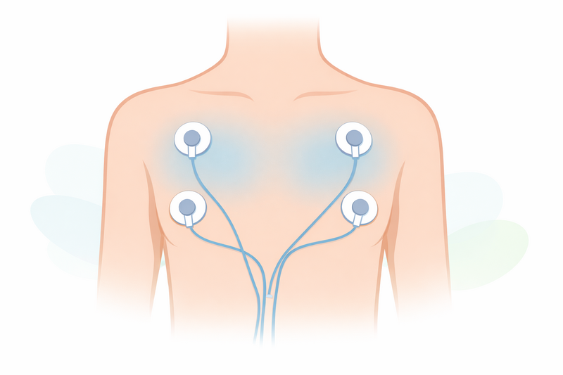
What happens during the test
Many people feel nervous about having an ECG, but the procedure is simple, painless, and usually takes less than 10 minutes. Here’s what you can expect when you go for an ECG:
- Preparation: You’ll be asked to remove your upper clothing and lie down on a couch or examination table. Sometimes, small areas on your chest may be shaved or cleaned to help the electrodes stick properly.
- Electrode placement: A healthcare professional will attach small sticky sensors, called electrodes, to your chest, arms, and legs. These electrodes are connected to the ECG machine by flexible wires.
- Recording the heart’s activity: Once the electrodes are in place, you’ll be asked to lie still and breathe normally for a short period. The machine will record the electrical signals produced by your heart from several different angles.
- Completion: After the recording is finished, the electrodes are removed, and you can get dressed and carry on with your day as usual. There are no restrictions after the test.
The test does not send any electricity into your body. It simply records the natural electrical signals created by your heart. The results are printed out as a series of lines and waves on paper or a screen, which your doctor will then interpret.
For some patients, a standard ECG is not enough to capture irregularities that happen occasionally. In these cases, your doctor may recommend a longer-term monitoring device, such as a Holter monitor, which you wear at home for 24 hours or more.
What ECGs can detect
ECGs can provide a wide range of information about the health of your heart. By analysing the patterns and timings of the electrical signals, doctors can detect various conditions and guide further management.
- Heart rhythm problems (arrhythmias): ECGs can identify if your heart is beating too fast (tachycardia), too slow (bradycardia), or irregularly (such as in atrial fibrillation).
- Heart attacks (myocardial infarction): Certain changes in the ECG pattern can indicate that a heart attack is happening or has happened in the past.
- Reduced blood flow (ischaemia): ECGs can sometimes show when parts of the heart muscle are not getting enough oxygen, which may be a sign of coronary artery disease.
- Enlarged heart chambers: Changes in the ECG waves can suggest if parts of the heart are enlarged, which can occur in conditions like high blood pressure or heart valve disease.
- Electrolyte imbalances: Abnormalities in the blood, such as potassium or calcium levels, can affect the heart’s electrical activity and show up on an ECG.
- Effects of medication: Some medicines can change the heart’s rhythm or conduction, and these changes are often visible on an ECG.
It’s important to remember that an abnormal ECG does not always mean you have a serious heart problem. Some changes are harmless, while others may require further investigation. Also, a normal ECG does not always rule out heart disease, especially if your symptoms come and go.
Doctors use ECGs as one piece of the puzzle, along with your medical history, symptoms, and other test results, to make a diagnosis or decide on treatment.
When further heart tests are needed
Sometimes, an ECG result raises questions that need to be explored with more specialised tests. Your doctor will decide if further investigations are necessary based on your symptoms, ECG findings, and overall risk factors.
- Ambulatory monitoring: If your symptoms are intermittent or your ECG is inconclusive, you may be asked to wear a portable monitor (like a Holter monitor or event recorder) for 24 hours or longer to capture your heart rhythm over time.
- Exercise (stress) ECG: This test records your heart’s activity while you walk on a treadmill or cycle, helping to detect problems that only occur during physical activity.
- Echocardiogram: This ultrasound scan shows pictures of your heart’s structure and function, giving more detail if an ECG suggests an abnormality.
- Cardiac MRI or CT scan: These imaging tests can provide detailed pictures of your heart and blood vessels if needed.
- Blood tests: In some cases, your doctor may check for markers of heart damage or look for electrolyte imbalances that could explain ECG changes.
Further tests are not always necessary. Sometimes, a single ECG is enough to reassure both you and your doctor. In other cases, ongoing monitoring or more in-depth investigations provide a clearer picture of your heart health.
If your doctor recommends more tests, it does not necessarily mean you have a serious problem. It often means they want to be thorough and ensure they have all the information they need to keep you safe and healthy.
If you are experiencing chest pain and are unsure whether it is an emergency or can wait, you may find helpful advice in our guide on when chest pain needs urgent attention versus when it is less serious.
In summary: An ECG is a valuable tool for detecting a range of heart issues and monitoring heart health. It is a quick, safe, and non-invasive test that forms a key part of cardiac assessment. While it can provide important clues about your heart’s function, it is only one part of the overall evaluation. If you have questions or concerns about your heart or your test results, always speak openly with your healthcare professional. They will guide you through the next steps, whatever they may be, and help you understand what your ECG truly means for your health and wellbeing.